Português
 English
TEMPORAL BONE BIOMODEL
Get to know the whole story, begun in the Otorhinolaryngology Service of Hospital das Clinicas in Sao Paulo
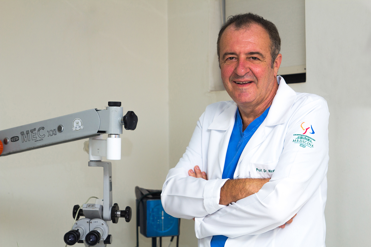
Prof Dr Ricardo Ferreira Bento
Otorhinolaryngologist
Titular Professor of Otorhinolaryngology at FMUSP.
“With 35 years of experience in teaching dissection courses and more than 150 courses held in Brazil and in 23 countries,
I was impressed with the similarity of this piece when compared to the cadaverous piece”
Know Auris-Otobone
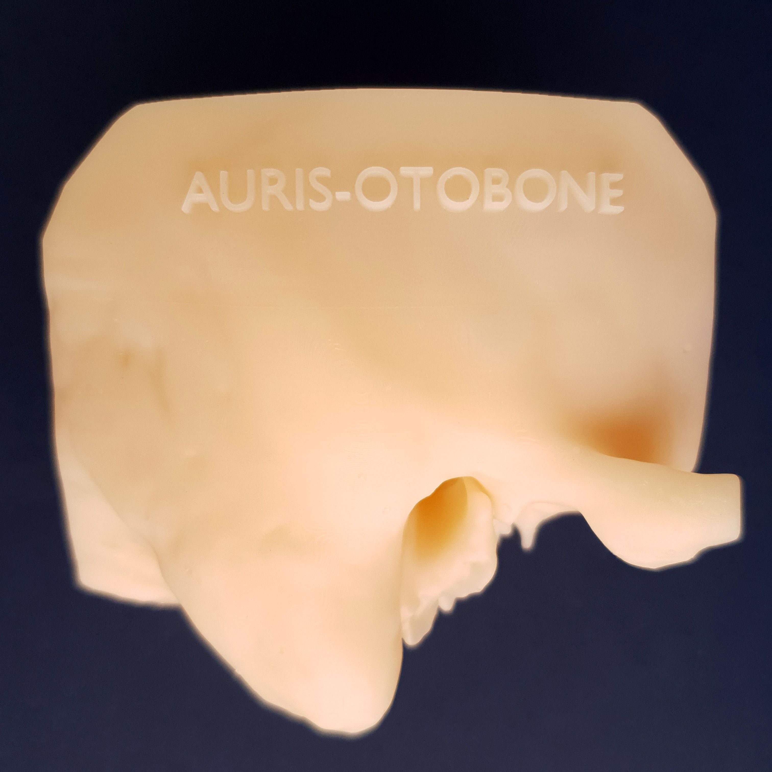
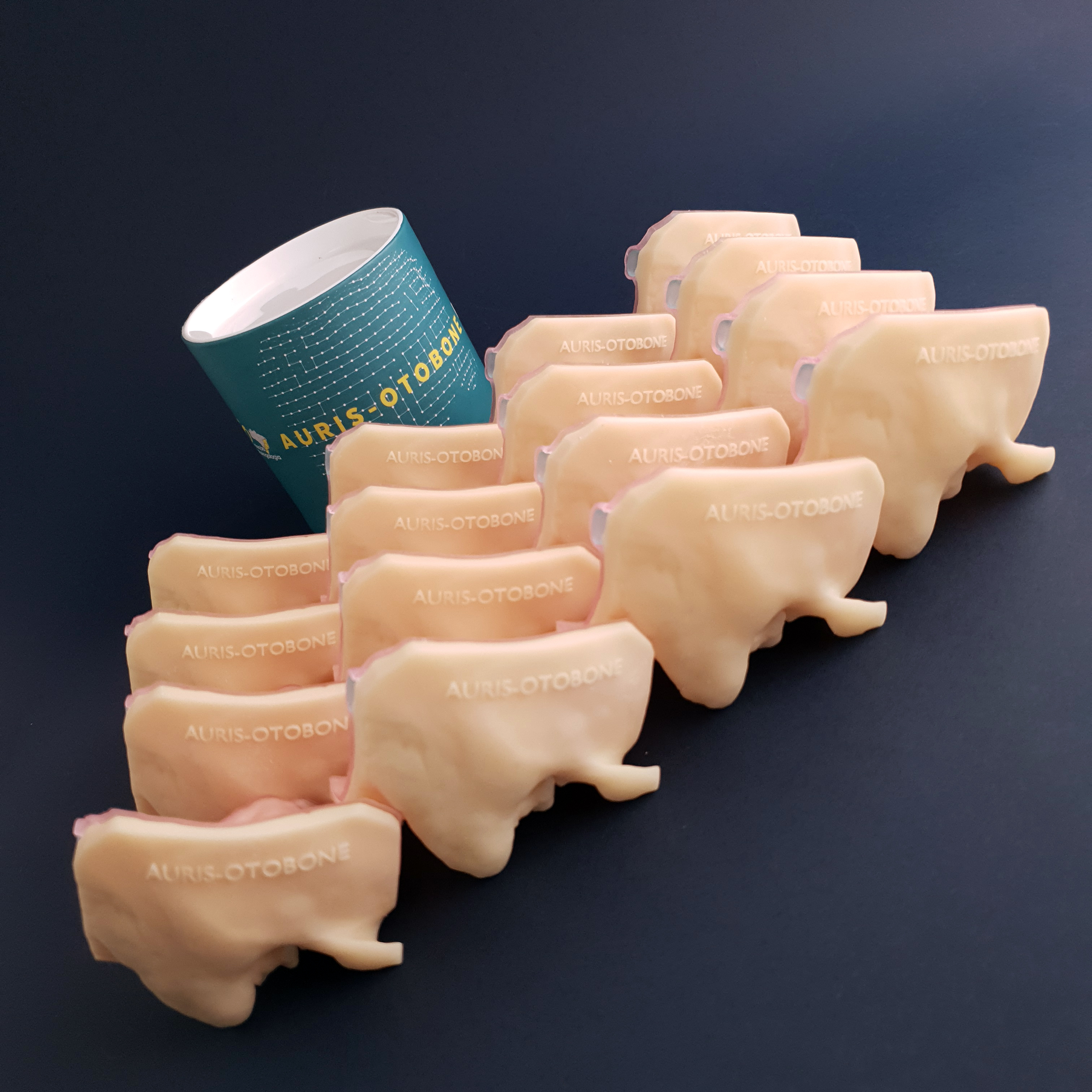
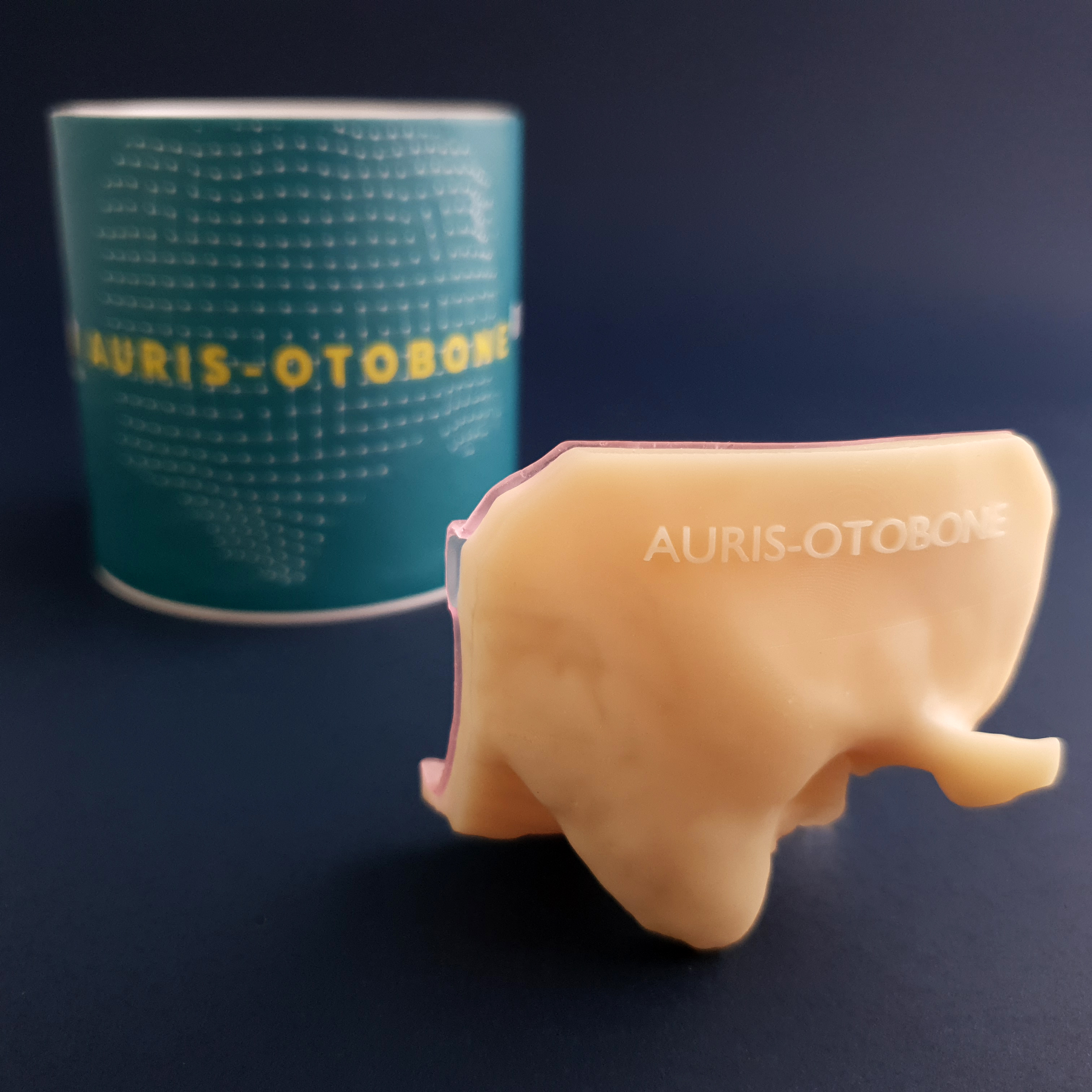
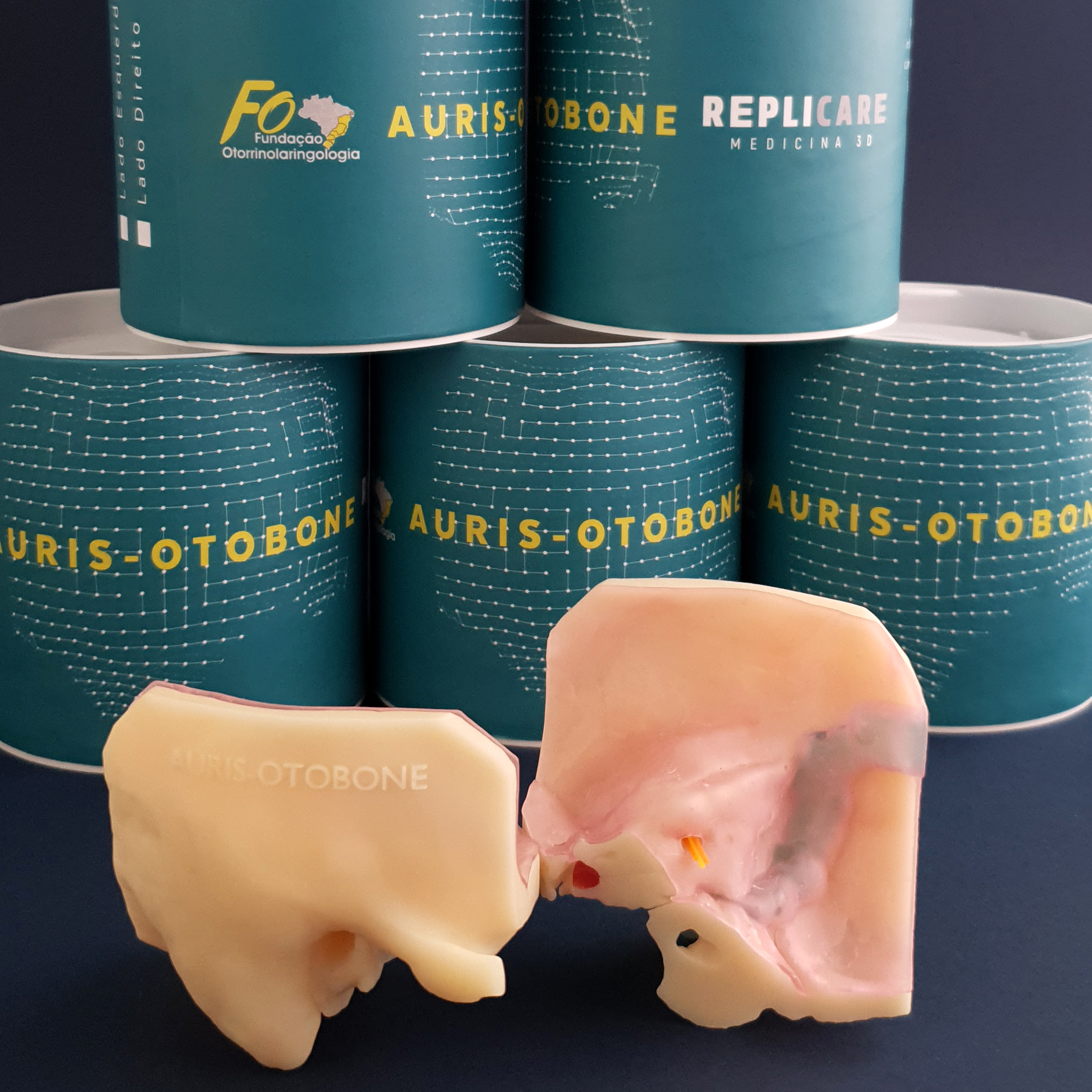
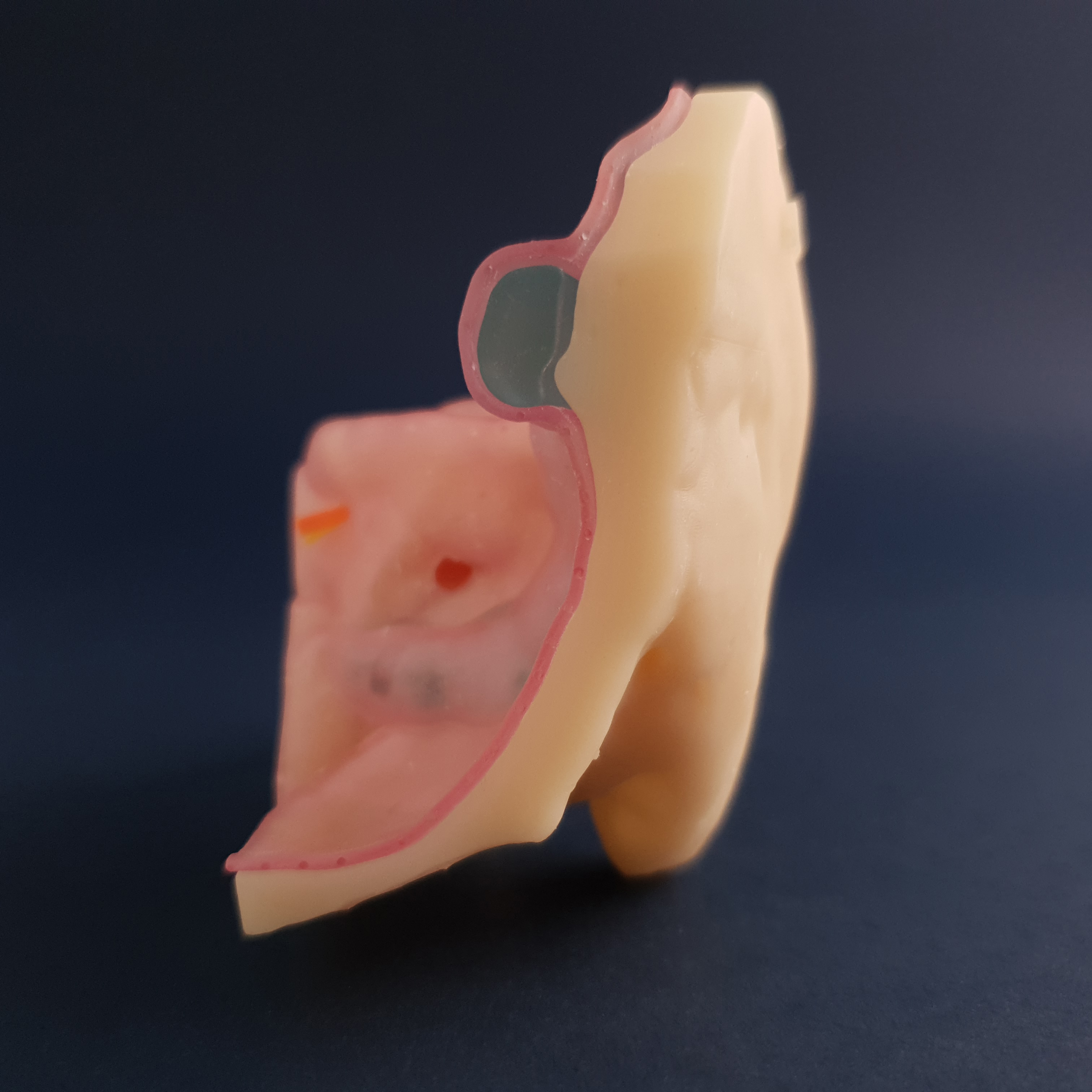
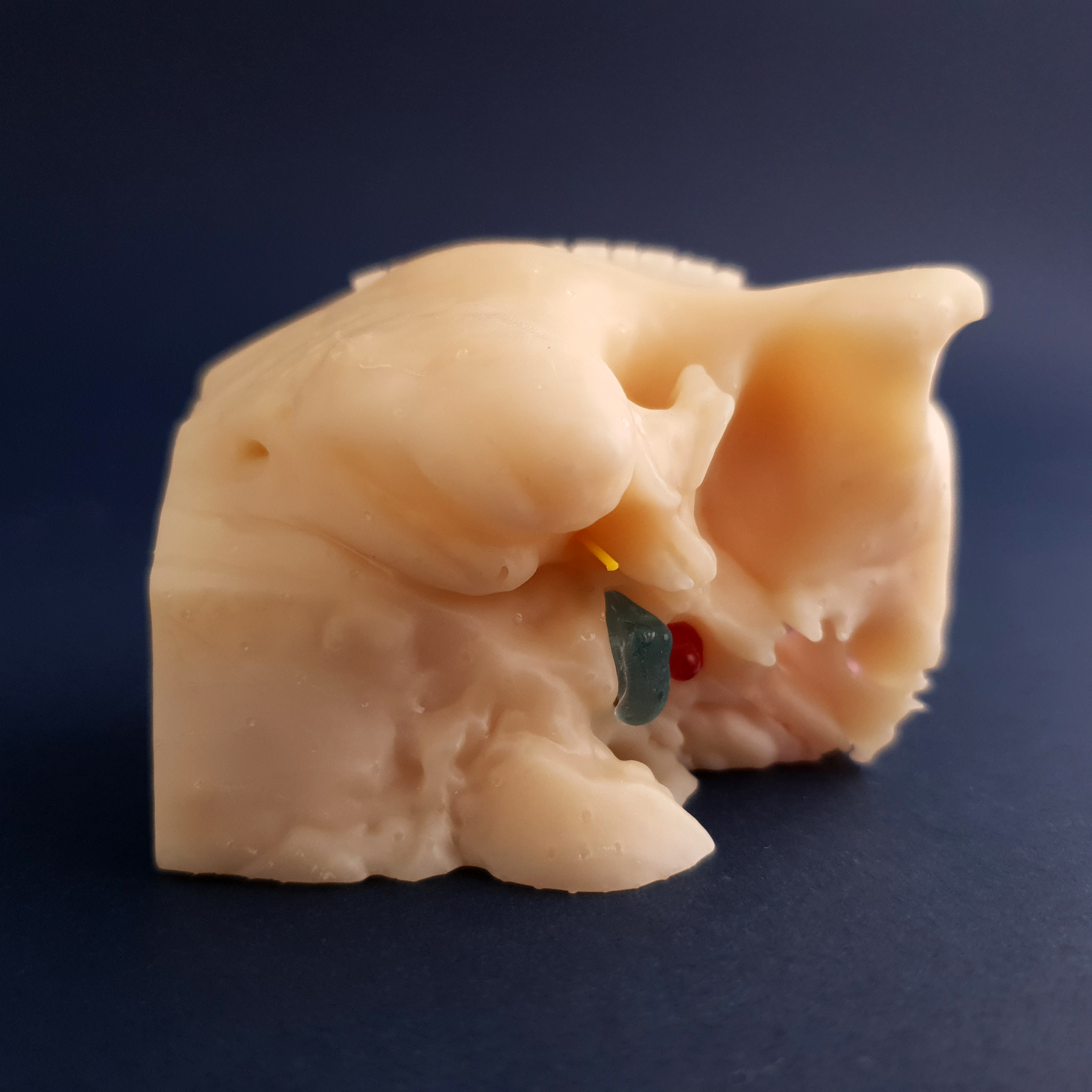
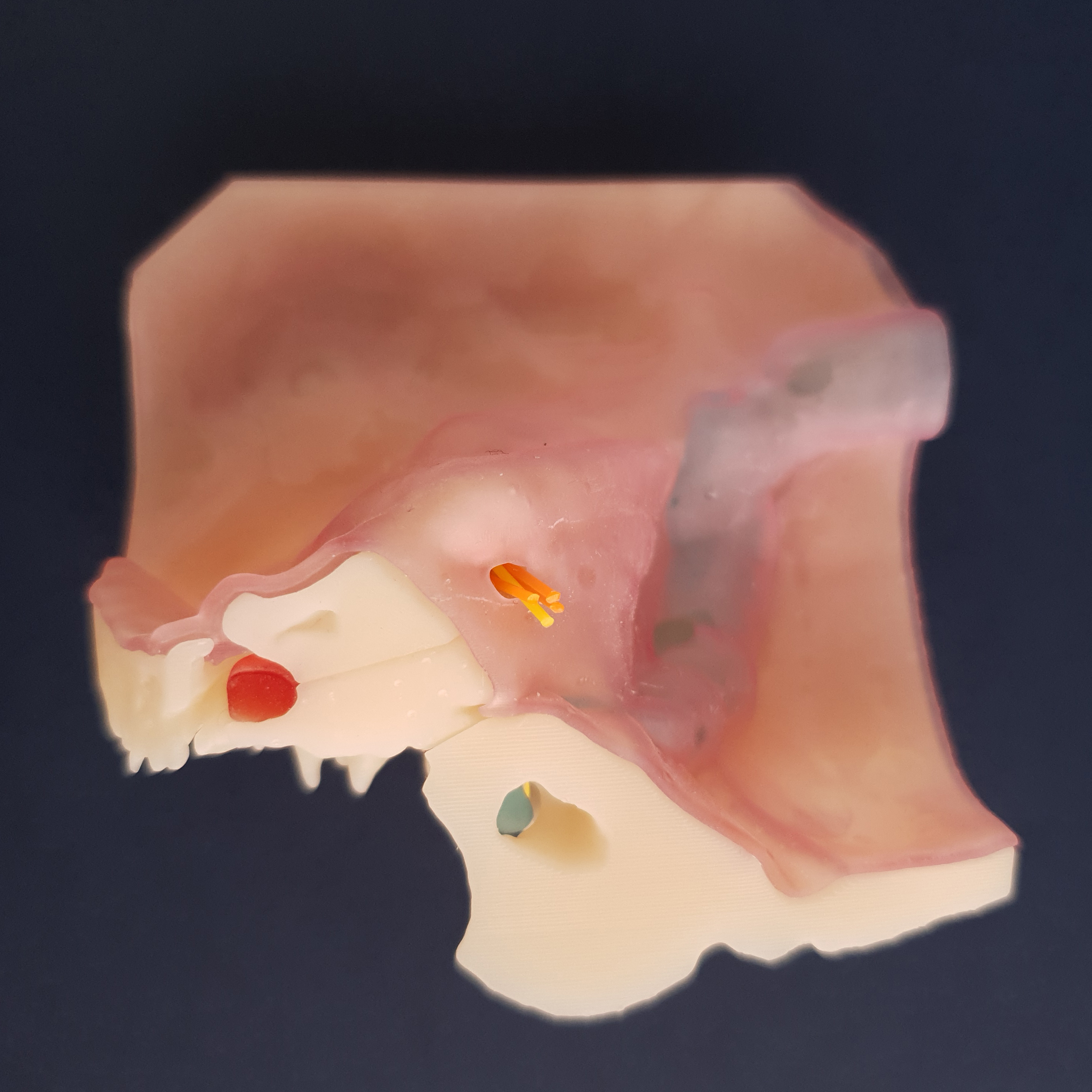
Auris Otobone® is an artificial temporal bone developed from a human CT, produced with a special material with density similar to a biological bone.
With difficulty in obtaining anatomical parts in corpses and with the increase in the number of specialists in educational institutes this is the ideal solution training.
Developed and tested in the laboratories of the medicine faculties of the USP and the Replicare-Medicina 3D
Currently available to doctors, educational institutions, students and companies for training implantable prostheses throughout Brazil and abroad.
Credits:
Prof. Dr Ricardo Ferreira Bento
Dra. Adriana Kosma Pires de Oliveira
Dra. Paula Tardim Lopes
Dr. Edson Leite Freitas
Dr. Fernando Balsalobre
Dr. Lucas Formighieri
Dr. Luiz Otávio de Mattos Coelho
Rafael Formighieri
PROVEN TECHNOLOGY AND BENEFITS
The physical and anatomical features, as well as the possibilities for its use were the object of study at 25 medical (fellows, assistant and residents of FMUSP and other services ent), after dissection, answered questionnaires anonymously.
It’s goal is help ear surgery training
IDEAL IN RESIDENT FORMATION
2. ldentify anatomical reference points: Facial Nerve, Auditory Tube, jugular vein, Mastoid notch of the digastric muscle, Antro, Attic, Ossicles, Oval and round window, Sigmoid sinus, Semicircular canals/ducts.
TRAINING IN EAR IMPLANTS
2. Anchored Prosthesis
IDEA IN UPDATING SPECIALIST'S KNOWLEDGE
2. Posterior Tympanotomy
3. Facial Nerve Decompression
4. Labyrinthectomy